

Product Introduction
|
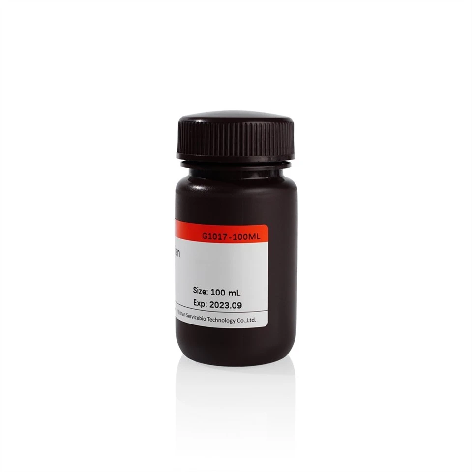
Product Name |
Cat.No. |
Spec. |
|
TTC Staining Solution |
G1017-100ML |
100 mL |
Product Description/Introduction
TTC (2, 3, 5-triphenyltetrazolium chloride), a lipid-soluble light-sensitive complex, can be used to detect seed viability or to detect ischemic infarction in mammalian tissues. The basic principle is that TTC is a proton acceptor of pyridine-nucleoside structural enzyme system in respiratory chain, and can react with dehydrogenase in normal tissues to form water insoluble red Triphenylformazan (TTF) compounds. Normal tissues and viable seeds are rich in dehydrogenase, which can react with TTC and appear red. If the tissue dies from ischemia or seed viability declines, it cannot react with TTC, and the tissue cannot be stained or the staining is very light. The degree of tissue ischemia infarction or seed vigor can be determined according to the staining site and depth.
The concentration of TTC in TTC staining solution of this product is 0.5%, which is suitable for the staining of ischemic infarction in animal tissues. The area of normal tissue is red, and the area of ischemic infarction is white.
Storage and Handling Conditions
Wet ice transportation; Store at 2-8℃ away from light, valid for 3 months.
Component
|
Component |
G1017-100ML |
|
TTC Staining Solution(0.5%,Animals) |
100 mL |
|
Product Manual |
|
Assay Protocol / Procedures
Sample Pretreatment: The ischemic infarcted tissues were taken immediately after the animals were sacrificed and placed in a clean petri dish. The tissues were wrapped with OCT embedding agent (G6059 is recommended) and flash-frozen at -20℃ for 30-60 min. If frozen sections could not be prepared in time, they should be stored at -80℃. Before staining, the tissue should be thawed slightly at room temperature and then sliced into sections with a very sharp blade with a thickness of 2-3 mm. Ensure that the sections are neat.
Staining Steps
1. TTC staining solution was preheated at 37℃ for 30 min.
2. Put the tissue sections into a clean container (be careful not to make the tissue be squeezed), add TTC staining solution to soak the tissue, and put the container in a 37℃ water bath to avoid light for about 30 min. During the period, the tissue was gently shaken every 10 min to contact the staining solution evenly, and the staining situation was observed in time.
3. When the tissue appears dark red, pour out TTC dye solution (which can be recycled), and slowly add fixing solution (G1101 is recommended) into the container to soak the tissue to terminate the staining reaction. Samples should be stored away from light.
4. Before taking photos, remove the tissue, blot the fixation solution with filter paper, and take photos with camera as needed.
1. Quick sampling is required to ensure fresh samples and maximize enzyme activity.
2. The stained samples are easy to fade, so photos should be taken as soon as possible.
3. Wear a lab coat and disposable gloves during the operation.
For Research Use Only!


